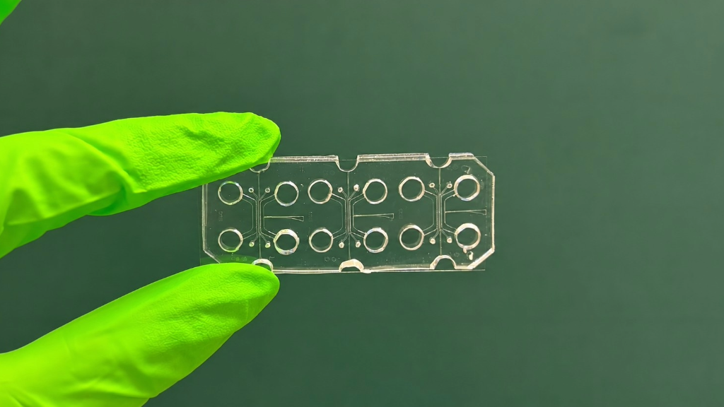
The platform, developed by researchers at Politecnico di Milano with Linköping University and Northeast Ohio Medical University, reproduces an innervated and vascularized carcinoma – The study was published in PNAS
Milan, 19 November 2026 – Testing drugs and studying the evolution of cancer cells by using a miniaturized experimental platform. A team from the Department of Electronics, Information and Bioengineering – DEIB at Politecnico di Milano, led by Professor Marco Rasponi, has contributed to the development of an organ-on-a-chip platform that reproduces an innervated and vascularized head and neck squamous cell carcinoma (HNSCC), using engineered extracellular matrices (eECMs), within a controlled human microenvironment.
The study underlying the project was published in the scientific journal PNAS – Proceedings of the National Academy of Sciences of the United States of America and was carried out together with researchers from Linköping University in Sweden, particularly the Laboratory of Molecular Materials (m2Lab), the Department of Otorhinolaryngology of Region Östergötland (Sweden), and Northeast Ohio Medical University. In the initial phase, patient-derived tumor cells from at Linköping University Hospital, cancer-associated fibroblasts and endothelial cells were cultured within a specially designed extracellular matrix. This consists of a functionalized hyaluronic acid-based gel “that allows cells to adhere to and remodel it, and, in the case of cancer cells, to form three-dimensional aggregates that simulate small tumor masses,” explained Marco Rasponi, coordinator of the project at Politecnico di Milano. His research group at the MiMic Lab was responsible for developing the engineered model, while their Swedish counterparts developed the biological component of the project. The first author of the study is Alessia Maria Pignataro, a former Politecnico di Milano student and now a PhD candidate at Linköping: the work originated from her master’s thesis at the MiMic Lab. The authors also include Alessandro Cordiale, a PhD candidate in the research group.
“We designed a particularly complex chip, because these organ-on-a-chip models normally work with one or two cell types, whereas in this case we had four: cancer cells, fibroblasts, endothelial cells and sensory neurons, each of which requires its own conditions to survive,” Rasponi pointed out. “We therefore worked in three stages: first on the endothelial and tumor components, then on the neural component, and finally on combining them, optimizing the individual cell populations. Based on the characterization tests of each cell type, we then developed the complete integrated model, which was used to conduct the study on the vascularized and innervated tumor.”
The final organ-on-a-chip makes it possible to understand how head and neck cancer – the sixth most common cancer worldwide – develops and evolves, and how it interacts with blood vessels and nerves, taking a further step towards precision and personalized medicine. “In the future, this could allow us to create different organ-on-a-chip models for an individual patient, on which to test the various available therapies and determine which are most effective,” Rasponi emphasized. The model also makes it possible to study the mechanisms of cancer pain, thanks to the presence of innervation, and to develop advanced therapies based on the therapeutic responses obtained, thereby providing biomedical research with a valid alternative to animal experimentation.
https://www.pnas.org/doi/10.1073/pnas.2602541123